Reichert’s Cartilage

  • Reichert’s cartilage is a cartilaginous structure that arises from the second pharyngeal arch.1
  • It it comprised of a cranial and caudal segment that give rise to different structures in the head and neck.2
    • The cranial segment of Reichert’s cartilage gives rise to the stapes, styloid process, and stylohyoid ligament.
    • The caudal segment gives rise to the lesser horns and upper part of the hyoid bone.
  • A similar structure is Meckel’s Cartilage, which arises from the first pharyngeal arch and gives rise to the malleus and incus.
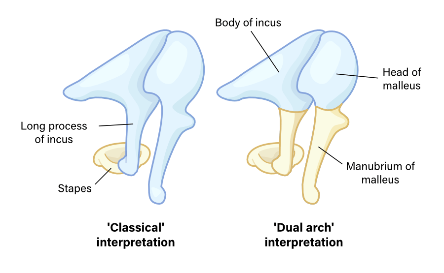
  • There have historically been two theories in the literature as to how the ossicles develop from Meckel’s cartilage and Reichert’s cartilage.3
    • The classical interpretation is that Meckel’s cartilage gives rise to the malleus and incus, while Reichert’s cartilage gives rise to the stapes, as described above.
    • The dual-arch interpretation is that Meckel’s cartilage gives rise to the epitympanic ossicles (such as the head of the malleus and the body of the incus), while Reichert’s cartilage gives rise to the mesotympanic ossicles (such as the manubrium of the malleus and the long and lenticular process of the incus).
  • Studies suggest that the classical interpretation is the more likely theory.

Theories of development from Meckel's Cartilage and Reichert's Cartilage

  1. Sadler, Thomas W. Langman’s medical embryology. Lippincott Williams & Wilkins, 2018. 

  2. Rodríguez‐Vázquez, J. F., et al. “Morphogenesis of the second pharyngeal arch cartilage (Reichert’s cartilage) in human embryos.” Journal of anatomy 208.2 (2006): 179-189. 

  3. Burford, Charlotte M., and Matthew J. Mason. “Early development of the malleus and incus in humans.” Journal of Anatomy 229.6 (2016): 857-870. 

Last updated October 27, 2022