Cutaneous Melanoma

  • This staging system only applies to melanoma involving the cutaneous skin.
  • Mucosal melanoma involving the head and neck, conjunctival melanoma, and uveal melanoma each have separate staging systems.
  • Mucosal melanoma involving the urethra/vagina/rectum/anus does not have an AJCC staging system.

Changes from the AJCC 7th Edition

T-Categorization

  • Mitotic rate is no longer part of the T categorization, but is still considered an important prognostic factor.
  • Tumor thickness measurements now are recorded to the nearest 0.1 mm, not the nearest 0.01 mm (because of the imprecision of measuring 0.01 mm differences)
  • T1a is now sub-divided by thickness (< 0.8 mm and 0.8 – 1.0 mm) in addition to the presence/absence of ulceration.

N-Categorization

  • The terms clinically occult and clinically detectable replaced the old terms microscopic and macroscopic nodal metastasis.
  • Non nodal regional metastases (microsatellites/satellite/in-transit metastases) are now stratified into N1c, N2c, or N3c based on the number of nodes involved.

M-Categorization

  • Distal metastases are now defined by both anatomic site and LDH level.
  • CNS metastasis is now placed in its own separate category (M1d) due to evidence that this is a particularly poor prognostic sign.

T-Categorization
T Category Criteria
TX
  • Primary tumour thickness cannot be assessed (eg. if the diagnosis was by curettage)
T0
  • No evidence of primary tumour (eg. unknown primary, or if the primary has completely regressed)
Tis
  • Melanoma in-situ (has not broken through basement membrane).
T1 ≤ 1.0 mm thickness
T1a
  • < 0.8 mm thickness without ulceration
T1b
  • 0.8 – 1.0 mm thickness with or without ulceration
  • OR < 0.8 mm thickness with ulceration
T2 > 1.0 – 2.0 mm thickness
T2a
  • > 1.0 – 2.0 mm thickness without ulceration
T2b
  • > 1.0 – 2.0 mm thickness with ulceration
T3 > 2.0 – 4.0 mm thickness
T3a
  • > 2.0 – 4.0 mm thickness without ulceration
T3b
  • > 2.0 – 4.0 mm thickness with ulceration
T4 > 4.0 mm thickness
T4a
  • > 4.0 mm thickness without ulceration
T4b
  • > 4.0 mm thickness with ulceration

General Rules

  • Thickness thresholds are ≤ 1.0 mm for T1, > 1.0 mm to 2.0 mm for T2, > 2.0 to 4.0 mm for T3, and > 4.0 mm for T4.
  • The presence of ulceration automatically upstages you to the 'b' sub-category.
  • New in the AJCC 8th Edition, T1 is now subdivided into T1a and T1b with a 0.8 mm cutoff, in addition to ulceration.
  • Only tumours that are < 0.8 mm without ulceration are considered T1a. Otherwise (if there is ulceration, or if it's 0.8 to 1.0 mm), it's considered T1b.

Tumour Thickness

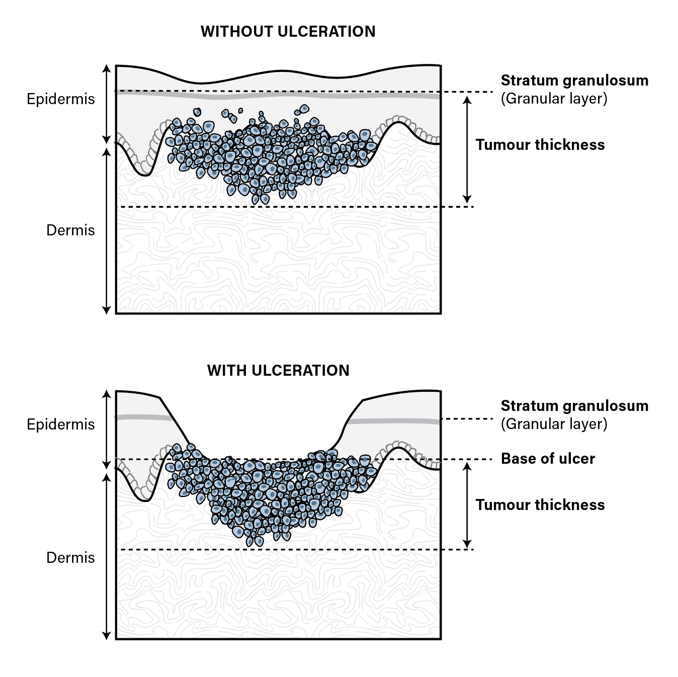
  • In non-ulcerated tumours, the thickness is measured from the granular layer of the epidermis (aka. the stratum granulosum).
  • In ulcerated tumours, the thickness is measured from the base of the ulcer.
  • Ulceration when characterizing a melanoma requires:
    1. Fullā€thickness epidermal defect.
    2. Reactive changes (such as fibrin deposition and the presence of neutrophils).
    3. Changes to the surrounding epidermis. These include thinning, effacement, or reactive hyperplasia in the absence of known trauma.

N-Categorization
N Category Criteria
NX
  • Regional nodes not assessed (eg. if SLN biopsy not done).
  • Exception: Thin melanomas (pT1) with no clinical evidence of distant metastases (cM0), can be considered cN0 due to their low incidence of metastases.
N0
  • No regional metastases detected
N1
N1a
  • 1 clinically occult lymph node
N1b
  • 1 clinically detected lymph node
N1c
  • 0 lymph nodes involved
  • AND the presence of microsatellites / satellites / in-transit metastases
N2
N2a
  • 2-3 clinically occult lymph nodes
N2b
  • 2-3 (with at least 1 clinically detected) lymph nodes
N2c
  • 1 lymph node (occult or detected)
  • AND the presence of microsatellites / satellites / in-transit metastases
N3
N3a
  • 4 or more clinically occult lymph nodes
N3b
  • 4 or more (with at least 1 clinically detected) lymph nodes
N3c
  • 2 or more lymph nodes (occult or detected)
  • AND the presence of microsatellites / satellites / in-transit metastases
  • OR the presence of any matted lymph nodes

General Rules

  • Generally ordered by the number of lymph nodes involved.
  • 1 lymph node involved places you in N1, 2-3 places you in N2, 4 or more places you into N3 (with exceptions for microsatellite/satellite/in-transit metastases).
    • If all of these lymph nodes are occult (ie. identified on Pathology only), then they are categorized with an a sub-category (N1a, N2a, N3a)
    • If any one of these lymph nodes are clinically detected (ie. seen clinically or radiologically), then they are categorized with a b sub-category (N1b, N2b, N3b).
    • Any microsatellite/satellite/in-transit metastases upstages you to the next numeric N category and is sub-categorized with a c (N1c, N2c, N3c). For instance, if you had 1 involved lymph node but satellite metastases, you would be categorized as an N2c (not N1).
    • Any matted lymph nodes (2 or more nodes that are adherent to one another on pathologic examination) brings you to the highest nodal sub-category (N3c).

Definitions

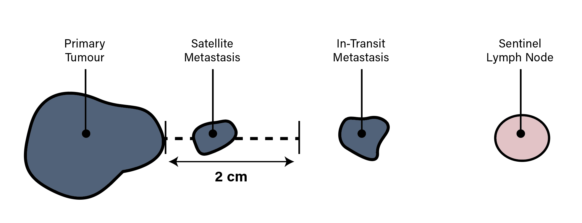
  • Microsatelitte Metastasis: cutaneous or subcutaneous metastases found adjacent to a primary melanoma on microscopic evaluation only (not clinically appreciable).
    • These tumour cells must be discontinuous from the primary tumour but cannot be separated only by fibrosis or inflammation (which may signify regression of the primary).
  • Satelitte Metastasis: Clinically appreciable cutaneous or subcutaneous metastases within 2 cm of the primary melanoma
  • In-Transit Metastasis: Clinically appreciable cutaneous or subcutaneous metastases > 2 cm away from the primary melanoma but not yet to the draining nodal basin.
  • Matted lymph nodes: Two or more nodes that adhere to one another, identified at the time the specimen is examined macroscopically on Pathology.

M-Categorization
M Category Criteria
M0

No evidence of distal metatasis

M1 Evidence of distal metastasis
M1a

Non-visceral metastasis (including to skin, soft tissue including muscle, and/or nonregional lymph nodes)

M1a(0)
  • Non-visceral metastasis with a normal LDH
M1a(1)
  • Non-visceral metastasis with an elevated LDH
M1b Lung metastasis
M1b(0)
  • Lung metastasis with a normal LDH
M1b(1)
  • Lung metastasis with an elevated LDH
M1c Visceral metastasis (excluding lung and CNS)
M1c(0)
  • Visceral metastasis with a normal LDH
M1c(1)
  • Visceral metastasis with an elevated LDH
M1d CNS metastasis
M1d(0)
  • CNS metastasis with a normal LDH
M1d(1)
  • CNS metastasis with an elevated LDH

General Rules

  • M-Categorization is divided by both anatomic site (M1a, M1b, M1c, or M1d) and by LDH level (assigned a (0) if normal or a (1) if elevated).
  • There are 4 different anatomic sites that are considered:
    1. Non-visceral metastasis classifies you as M1a.
    2. Lung metastasis classifies you as M1b.
    3. Visceral metastasis classifies you as M1c.
    4. CNS metastasis classifies you as M1d.
  • The LDH level is factored in independently, and adds a (0) or (1) modifier if normal or elevated respectively.
    • For instance, hepatic metastasis with a normal LDH would be assigned M1c(0), while lung metastasis with an elevated LDH would be assigned M1b(1).

Group Staging

Clinical Group Staging

N0
M0
N1-3
M0
Any N
M1
Tis 0 III IV
T1a IA III IV
T1b IB III IV
T2a IB III IV
T2b IIA III IV
T3a IIA III IV
T3b IIB III IV
T4a IIB III IV
T4b IIC III IV

General Rules

  • Any nodal metastasis upstages to stage III
  • Any distal metastasis upstages to stage IV
  • In the absence of nodal metastasis or distal metastasis your group stage is either I or II:
    • Stage I is subdivided into IA/IB.
    • Stage II is subdivided into IIA/IIB/IIC.
  • Only Tis, N0, M0 is classified as Stage 0
  • Only T1a, N0, M0 is classified as Stage IA
  • Only T4b, N0, M0 is classified as Stage IIC
  • Every 2 consecutive T-substages after T1a and before T4b is given the same group stage.
    • For instance (T1b, N0, M0) and (T2a, N0, M0) are both staged as IB, while (T2b, N0, M0) and (T3a, N0, M0) are both staged as IIA.

Pathologic Group Staging

N0
M0
N1a
M0
N1b
M0
N1c
M0
N2a
M0
N2b
M0
N2c
M0
N3a
M0
N3b
M0
N3c
M0
M1
T0 IIIB IIIB IIIC IIIC IIIC IIIC IV
T1a Ia IIIA IIIB IIIB IIIA IIIB IIIC IIIC IIIC IIIC IV
T1b Ib IIIA IIIB IIIB IIIA IIIB IIIC IIIC IIIC IIIC IV
T2a Ib IIIA IIIB IIIB IIIA IIIB IIIC IIIC IIIC IIIC IV
T2b IIa IIIB IIIB IIIB IIIB IIIB IIIC IIIC IIIC IIIC IV
T3a IIa IIIB IIIB IIIB IIIB IIIB IIIC IIIC IIIC IIIC IV
T3b IIb IIIC IIIC IIIC IIIC IIIC IIIC IIIC IIIC IIIC IV
T4a IIb IIIC IIIC IIIC IIIC IIIC IIIC IIIC IIIC IIIC IV
T4b IIc IIIC IIIC IIIC IIIC IIIC IIIC IIID IIID IIID IV

General Rules

  • Same general concepts as the clinical staging system, in that:
    • Any nodal metastasis upstages to stage III
    • Any distal metastasis upstages to stage IV
    • Group staging for N0, M0 disease is the same as the clinical staging.
  • The only difference with pathological staging is that stage III is subdivided into 4 groups (IIIa/IIIb/IIIc/IIId) depending on the primary tumour and nodal status (see above).