| T Category | Criteria |
|---|---|
| TX |
Primary tumour cannot be assessed |
| Tis |
Carcinoma in situ. |
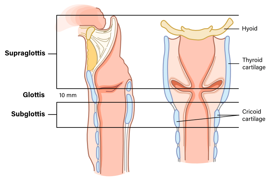
| T1 |
Tumour limited to 1 subsite of the supraglottis only and with normal vocal fold mobility. |
| T2 |
|
| T3 |
|
| T4 | Moderately advanced (T4a) or very advanced (T4b) disease |
| T4a |
|
| T4b |
|
| T Category | Criteria |
|---|---|
| TX |
Primary tumour cannot be assessed |
| Tis |
Carcinoma in situ. |
| T1 | Tumour limited to the vocal folds only. |
| T1a |
Tumour limited to one vocal fold only. |
| T1b |
Tumour involves both vocal folds (eg. involving the anterior commissure). |
| T2 |
|
| T3 |
|
| T4 | Moderately advanced (T4a) or very advanced (T4b) disease |
| T4a |
|
| T4b |
|
| T Category | Criteria |
|---|---|
| TX |
Primary tumour cannot be assessed |
| Tis |
Carcinoma in situ. |
| T1 |
Tumour is limited to the subglottis. |
| T2 |
|
| T3 |
|
| T4 | Moderately advanced (T4a) or very advanced (T4b) disease |
| T4a |
|
| T4b |
|
| N Category | Criteria |
|---|---|
| NX |
Regional lymph nodes cannot be assessed. |
| N0 |
No regional lymph node metastasis. |
| N1 |
|
| N2 | |
| N2a |
|
| N2b |
|
| N2c |
|
| N3 | |
| N3a |
Metastasis in any lymph node > 6 cm without extranodal extension |
| N3b |
Metastasis in any lymph node with clinically overt extranodal extension. |
| N Category | Criteria |
|---|---|
| NX |
Regional lymph nodes cannot be assessed. |
| N0 |
No regional lymph node metastasis. |
| N1 |
|
| N2 | |
| N2a |
|
|
|
| N2b |
|
| N2c |
|
| N3 | |
| N3a |
Metastasis in any lymph node > 6 cm without extranodal extension |
| N3b |
|
|
|
| M Category | Criteria |
|---|---|
| M0 | No distant metastasis |
| M1 | Distant metastasis present |
| N0 M0 |
N1 M0 |
N2 M0 |
N3 M0 |
Any N M1 |
|
| T1 | I | III | IVA | IVB | IVC |
| T2 | II | III | IVA | IVB | IVC |
| T3 | III | III | IVA | IVB | IVC |
| T4a | IVA | IVA | IVA | IVB | IVC |
| T4b | IVB | IVB | IVB | IVB | IVC |