Oral Cavity

Changes from the AJCC 7th Edition

T-Categorization

  • The AJCC 8th Edition was revised to exclude the dry (vermillion) lip from the oral cavity.
  • In previous versions cancers of the dry lip were considered part of the oral cavity. Now they are staged as cutaneous carcinomas of the head and neck.
  • In addition, revisions to the 8th Edition Guidelines in June of 2018 changed the T-staging for the oral cavity significantly (see below).
  • Depth of invasion now plays a significant role in T-categorization in addition to tumour size.
  • Extrinsic tongue muscle invasion no longer upcategorizes to T4 because this is now encapsulated in the depth of invasion.

N-Categorization

  • Uses the new shared nodal staging system described previously.

T-Categorization
T Category Criteria
TX

Primary tumour cannot be assessed

Tis

Carcinoma in situ.

T1
  • Tumour 2 cm or smaller in greatest dimension.</span>
  • AND DOI ≤ 5 mm
T2
  • Tumour > 2 and ≤ 4 cm in greatest dimension with
    DOI ≤ 10 mm
  • OR tumour 2 cm or smaller in greatest dimension with DOI > 5 mm but ≤ 10 mm.
T3
  • Tumour > 2cm and ≤ 4cm with DOI > 10 mm
  • OR tumour > 4 cm with DOI ≤ 10 mm
T4 Moderately advanced (T4a) or very advanced (T4b) disease
T4a
  • Tumour > 4 cm AND DOI > 10 mm
  • OR tumour invasion of adjacent structures only (including through cortical bone of the mandible or maxilla, or involves the maxillary sinus or skin of the face).
  • Note that superficial erosion of a bone/tooth socket (alone) by a gingival primary is not sufficient to classify a tumor as T4.
T4b
  • Very advanced local disease
  • Tumour invasion of the masticator space, pterygoid plates, skull base, or encasement of the internal carotid.

General Rules

  • Previously, T1, T2, and T3 were defined only by tumour size (≤2 cm, >2 - 4 cm, and >4 cm respectively). Now depth of invasion (≤5 mm, >5 - 10 mm, and >10 mm) plays a role as well.
  • To be considered T1 you must have a tumour size ≤ 2 cm AND a DOI ≤ 5 mm.
  • Increasing DOI essentially increases your T categorization by 1. For instance, if you have a ≤2 cm tumour but your DOI is 5-10 mm, then this is T2 not T1. If you have a a >2 - 4 cm tumour but your DOI is >10 mm then this is T3 and not T2. If you have a >4 cm tumour but your DOI is >10 mm then this is T4a and not T3.
  • Many online sources will contradict this because the 8th Edition had multiple revisions which significantly changed oral cavity staging. Not only do they not exclude the dry (vermillion) lip, but the definitions for T2/T3/T4a have changed as well compared to the initial release of the 8th Edition. See published changes below:

Changes in the 8th Edition 3rd Printing

AJCC 8th Edition Supplement

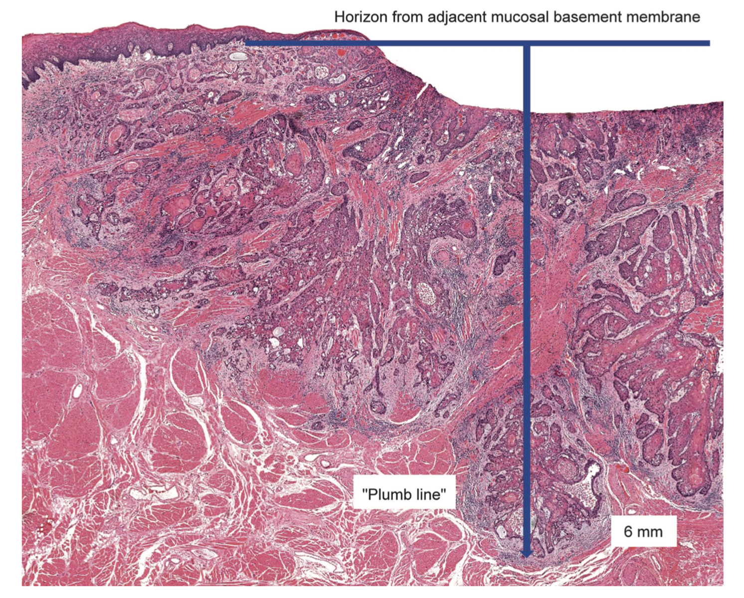
Depth of Invasion versus Tumour Thickness

  • Depth of Invasion (DOI) is not the same as tumour thickness.
  • Tumour thickness measures the distance from the most superficial to the deepest aspect of the tumour.
  • Depth of invasion is calculated by measuring the distance to the deepest aspect of the tumour measured from a line parallel to the basement membrane of the nearest intact squamous mucosa.
  • A plumb line is drawn from the 'horizon' from the nearest intact basement membrane and is used to measure the DOI.
  • If a tumour is very exophytic, the tumour thickness will be greater than the depth of invasion. Conversely, if a tumour is very ulcerated, the depth of invasion may be greater than the tumour thickness (as seen in the example below).

N-Categorization

Clinical Nodal Categorization
(Shared Categorization System)

N Category Criteria
NX

Regional lymph nodes cannot be assessed.

N0

No regional lymph node metastasis.

N1
  • Single ipsilateral lymph node that is 3 cm or less in maximal dimension without extranodal extension
N2
N2a
  • Single ipsilateral lymph node that is > 3 cm but ≤ 6 cm in maximal dimension without extranodal extension
N2b
  • Mulple ipsilateral lymph nodes that are ≤ 6 cm in maximal dimension without extranodal extension
N2c
  • Bilateral or contralateral lymph nodes that are ≤ 6 cm in maximal dimension without extranodal extension
N3
N3a

Metastasis in any lymph node > 6 cm without extranodal extension

N3b

Metastasis in any lymph node with clinically overt extranodal extension.

Pathologic Nodal Categorization
(Shared Categorization System)

N Category Criteria
NX

Regional lymph nodes cannot be assessed.

N0

No regional lymph node metastasis.

N1
  • Single ipsilateral lymph node that is 3 cm or less in maximal dimension without extranodal extension
N2
N2a
  • Single ipsilateral lymph node that is > 3 cm but ≤ 6 cm in maximal dimension without extranodal extension
  • OR metastasis to a single ipsilateral lymph node that is 3 cm or less in maximal dimension WITH extranodal extension
N2b
  • Mulple ipsilateral lymph nodes that are ≤ 6 cm in maximal dimension without extranodal extension
N2c
  • Bilateral or contralateral lymph nodes that are ≤ 6 cm in maximal dimension without extranodal extension
N3
N3a

Metastasis in any lymph node > 6 cm without extranodal extension

N3b
  • Metastasis in any lymph node with clinically overt extranodal extension.
  • EXCEPT if it a single ipsilateral node that is 3 cm or less with extranodal (which would instead be categorized as N2a as above).

General Rules

  • Uses the Shared Nodal Staging System described previously.
  • If a patient has any clinical extranodal extension, they are automatically be categorized in the maximum possible N sub-category (N3b).
  • Pathologic N categorization is almost the same except for the isolated situation of having a single ipsilateral lymph node that is 3 cm or less with ENE identified only on pathology.
    • Instead of being up-categorized to N3b, this specific case is up-categorized to N2a instead.
    • In all other situations where there is ENE (clinical or pathologic), they are still up-categorized to N3b.
  • Clinically the AJCC defines ENE as invasion of skin, infiltration of musculature/fixation to adjacent structures on clinical examination, cranial nerve, brachial plexus, sympathetic trunk or phrenic nerve invasion with dysfunction.

Extranodal Extension (ENE)

  • Pathologically, ENE is defined as extension of metastatic tumour beyond the confines of the lymph node itself.
  • Clinically the AJCC defines ENE as invasion of skin, infiltration of musculature/fixation to adjacent structures on clinical examination, cranial nerve, brachial plexus, sympathetic trunk or phrenic nerve invasion with dysfunction.

M-Categorization
M Category Criteria
M0

No distant metastasis

M1

Distant metastasis present


Group Staging

Shared Group Staging System

N0
M0
N1
M0
N2
M0
N3
M0
Any N
M1
T1 I III IVA IVB IVC
T2 II III IVA IVB IVC
T3 III III IVA IVB IVC
T4a IVA IVA IVA IVB IVC
T4b IVB IVB IVB IVB IVC

General Rules

  • Uses the Shared Group Staging System described previously.
  • Straightforward to remember due to the pattern seen above when creating a table against the T and N staging.
  • (T1 N0) is staged as Stage I, and (T2, N0) is staged as Stage II. Thereafter, each increasing stage is immediately to the right and below the previous stage.